Plenodomus tracheiphilus(DEUTTR)
Photos
For publication in journals, books or magazines, permission should be obtained from the original photographers with a copy to EPPO.
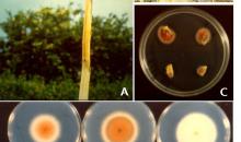
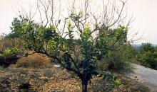
A Defoliation and dieback on a lemon twig. B Salmon-orange stain of the wood on the trunk of a lemon tree. The bark has been peeled off to show the discoloration of the wood. C Isolation of P. tracheiphila from an infected twig with the typical salmon-orange discoloration. Mycelium of a red-pigmented (chromogeneous) variant developing from wood pieces plated on PDA. D Colony morphology of 3 isolates on 2 media: Czapek-Dox (top lane) and PDA (bottom lane).
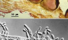
A Hand-made tangential section of a withered lemon twig showing pycnidia immersed in the cortex. Note the necks of pycnidia emerging through the epidermis. B Phialides and phialoconidia (differential interference contrast).
Courtesy: S. Grasso (IT).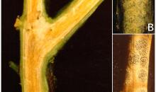
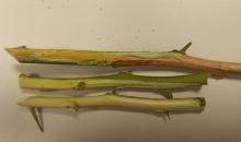
A Longitudinal section of a lemon twig showing the typical salmon-orange stain of the xylem. B Comparison between acervuli of C. gloeosporioides (top) and pycnidia of P. tracheiphila (bottom) on a 2-3 year old dried lemon twig. C Dried lemon twig with acervuli of C. gloeosporioides.
Courtesy: S. Grasso (IT).
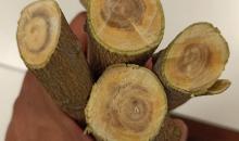
Wood discoloration in a lemon tree infected by mal secco.
Courtesy: G. Perrotta Università di Calabria (IT).
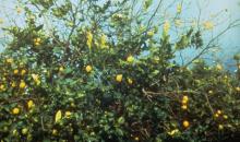
First symptoms in a branch of a lemon tree infected by Plenodomus tracheiphilus
Courtesy: Miguel Ángel Fernández, Plant Health Service. Autonomous Community Region of Murcia-Spain
