Melampsora medusae(MELMME)
Photos
For publication in journals, books or magazines, permission should be obtained from the original photographers with a copy to EPPO.

Urediniospores and paraphyses of Melampsora medusae f. sp. deltoidae observed by light microscopy. Note the smooth (non-echinulate) patches at the equator of the urediniospores, and the thin (3-6 µm) paraphyse wall.
Courtesy: Pascal Frey (INRAE, Nancy, France)
Spermogonia of M. medusae on Larix decidua, following inoculation of needles in the glasshouse with basidiospores.
Courtesy: A.L. Schippers Jr - USDA (US).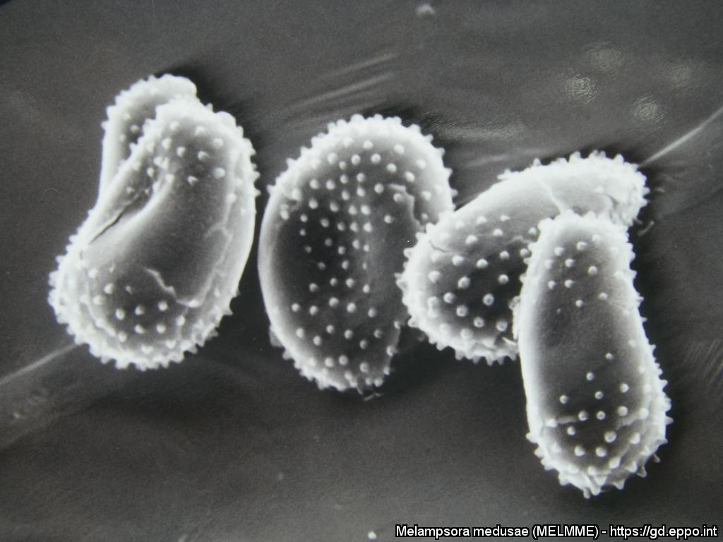
Melampsora medusae f. sp. deltoidae urediniospores observed by scanning electron microscopy. Note the smooth (non-echinulate) patches at the equator of the urediniospores
Courtesy: Pascal Frey (INRAE, Nancy, France)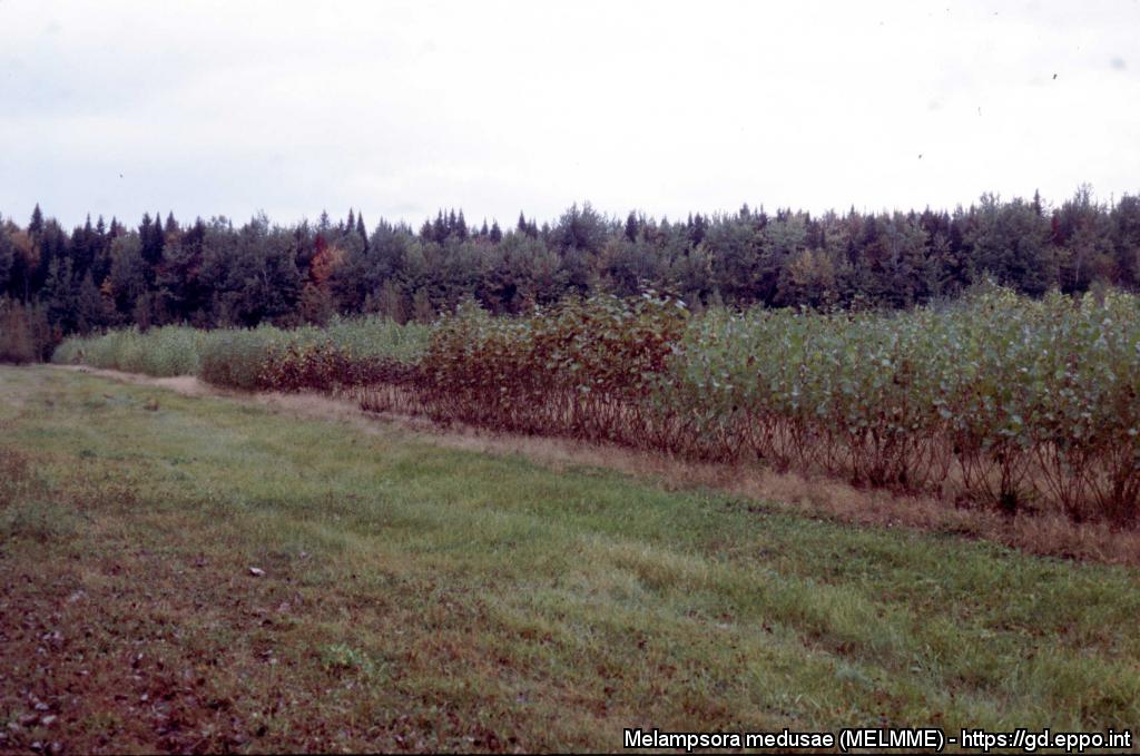
Poplar nursery trial, showing strong differencies in the susceptibility of poplar genotypes to Melampsora medusae f. sp. deltoidae (Pointe Platon, Lotbinière, Quebec, Canada)
Courtesy: Pascal Frey (INRAE, Nancy, France)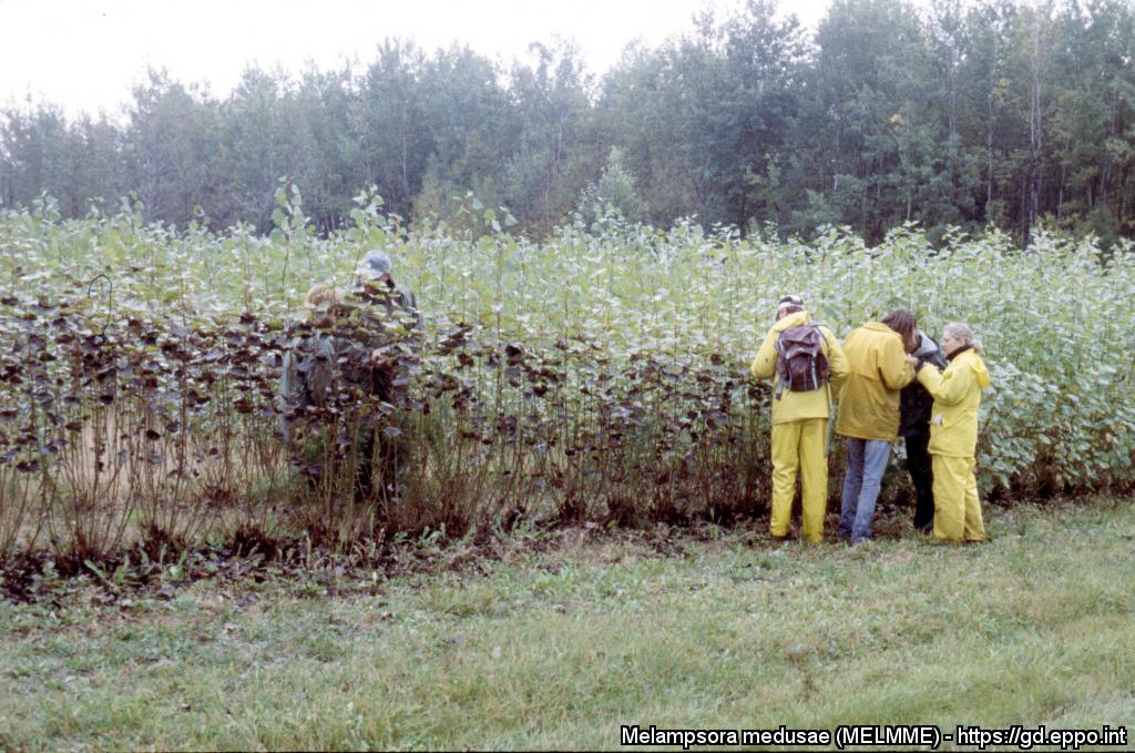
Poplar nursery trial, showing strong differencies in the susceptibility of poplar genotypes to Melampsora medusae f. sp. deltoidae (Pointe Platon, Lotbinière, Quebec, Canada)
Courtesy: Pascal Frey (INRAE, Nancy, France)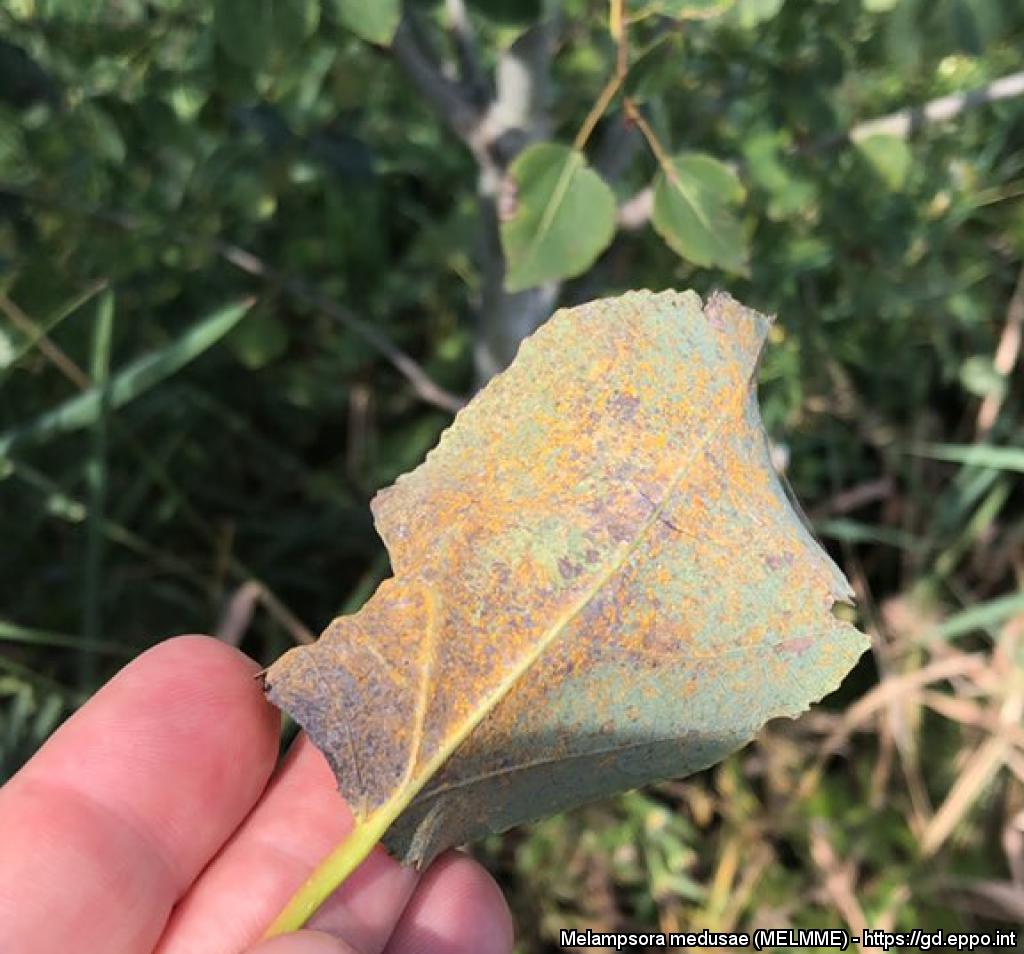
Uredinia of M. medusae f. sp. tremuloidae on the underside of leaves of aspen (Populus tremuloides).
Courtesy: Philippe Tanguay (Natural Resources Canada, Laurentian Forestry Centre, Canada)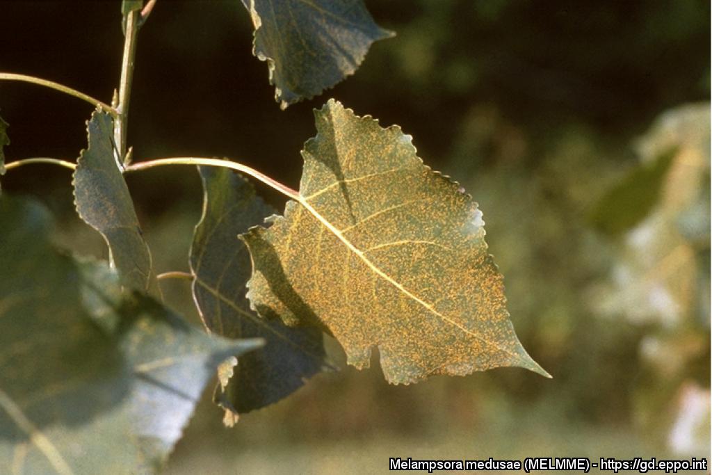
Yellowish uredinia of M. medusae on the underside of leaves of poplar (Populus deltoides).
Courtesy: A.L. Schippers Jr - USDA (US).