Phyllocoptes fructiphilus(PHYCFR)
Photos
All photos included on this page can only be used for educational purposes.
For publication in journals, books or magazines, permission should be obtained from the original photographers with a copy to EPPO.
For publication in journals, books or magazines, permission should be obtained from the original photographers with a copy to EPPO.

Light micrographs of prodorsal shield ornamentation for Callyntrotus schlechtendali, P. resovius, P. adalius and P. fructiphilus
Courtesy: Tobiasz Druciarek, University of Arkansas, Fayetteville, USA
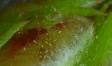
Various stages feeding on an unopened Rosa flower
Courtesy: Patrick Di Bello, Oregon State University (US)
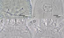
Qualitative characters of Phyllocoptes arcani: A – prodorsal shield, B – coxigenital region, and Phyllocoptes fructiphilus: C – prodorsal shield, D – coxigenital region.
Courtesy: Druciarek et al. 2021
Phyllocoptes from rose, antero-dorsal females: A – P. fructiphilus; B – P. arcani; C – P. adalius
Courtesy: Druciarek et al. 2021
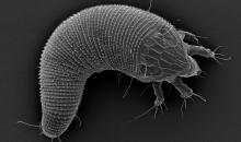
SEM pictures of Callyntrotus schlechtendali, Phyllocoptes resovius, P. adalius and P. fructiphilus. Note the characteristic waxy secretions present in case of C. schlechtendali and P. resovius (arrows). Such structures are absent in case of P. adalius and P. fructiphilus
Courtesy: Tobiasz Druciarek, University of Arkansas, Fayetteville, USA

