Xanthomonas arboricola pv. pruni(XANTPR)
Photos
For publication in journals, books or magazines, permission should be obtained from the original photographers with a copy to EPPO.
Spots on a plum fruit (Prunus domestica cv. Shiro).
Courtesy: U. Mazzucchi, Universita degli Studi, Bologna (IT).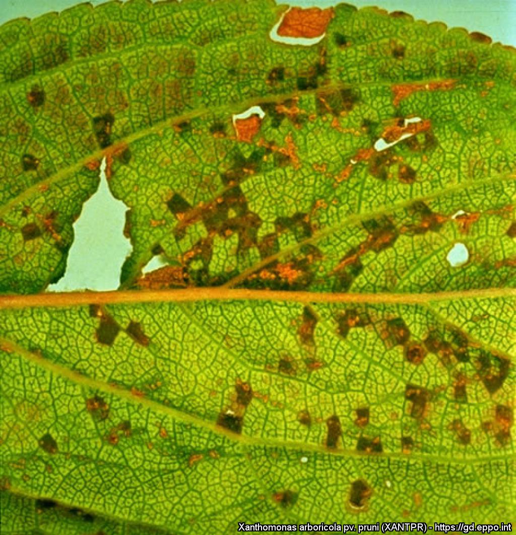
Old angular spots on a plum leaf (P. domestica cv. Calita).
Courtesy: U. Mazzucchi, Universita degli Studi, Bologna (IT).
Symptoms on peach fruit (P. persica).
Courtesy: U. Mazzucchi, Universita degli Studi, Bologna (IT).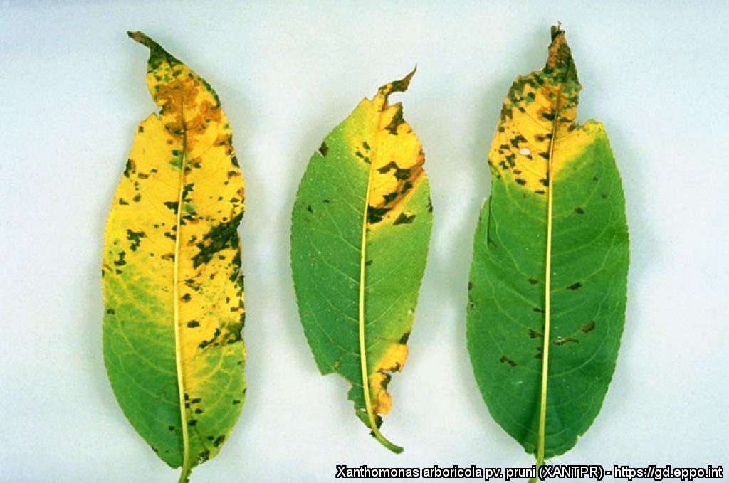
Peach leaves (P. persica cv. Maria Serena) with numerous spots.
Courtesy: U. Mazzucchi, Universita degli Studi, Bologna (IT).
Stem cankers on plum (P. domestica cv. Calita), after removal of the outer bark layer.
Courtesy: U. Mazzucchi, Universita degli Studi, Bologna (IT).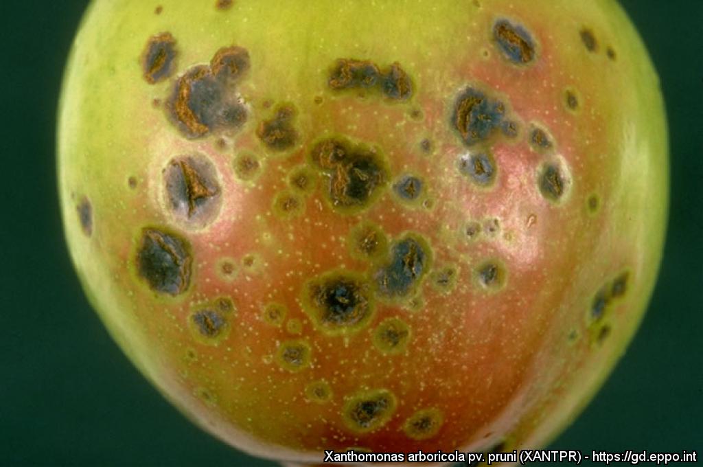
Spots on plum fruit (P. domestica cv. Frontier).
Courtesy: U. Mazzucchi, Universita degli Studi, Bologna (IT).
Spots caused by X. arboricola pv. pruni on two peach leaves (P. persica cv. Elegant Lady).
Courtesy: U. Mazzucchi, Universita degli Studi, Bologna (IT).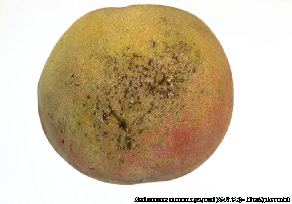
Small circular sunken brown lesions on peach fruit.
Courtesy: H.L. Keil (US).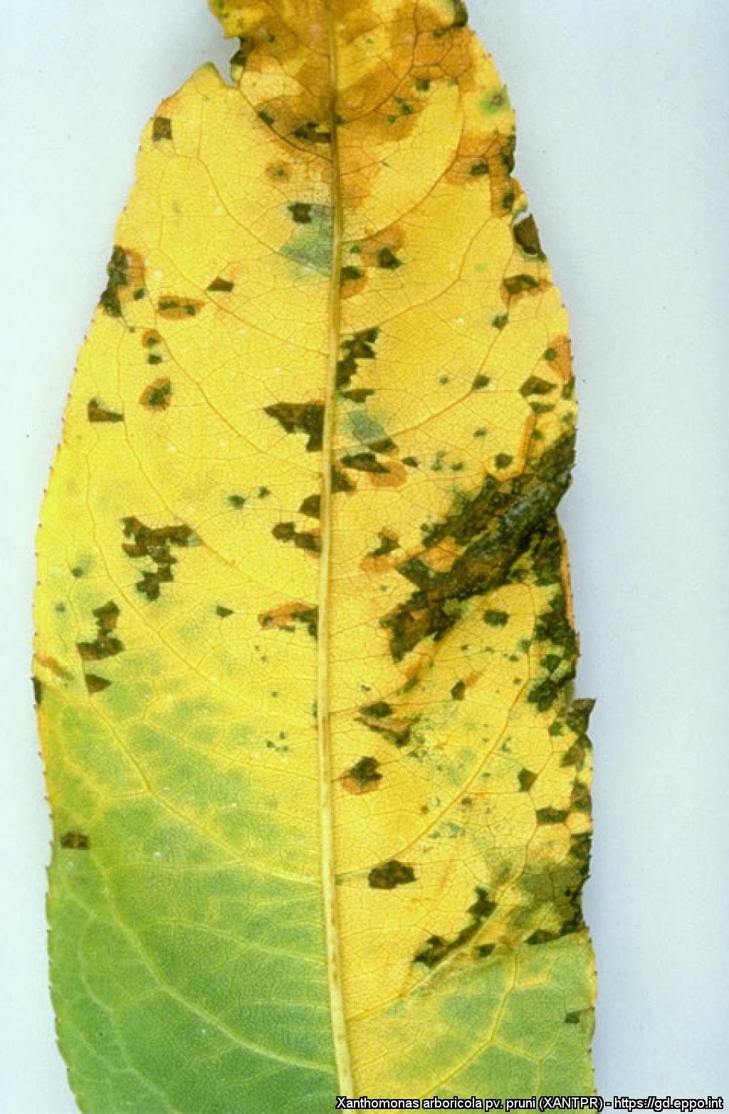
Detail of a peach leaf (P. persica cv. Maria Serena).
Courtesy: U. Mazzucchi, Universita degli Studi, Bologna (IT).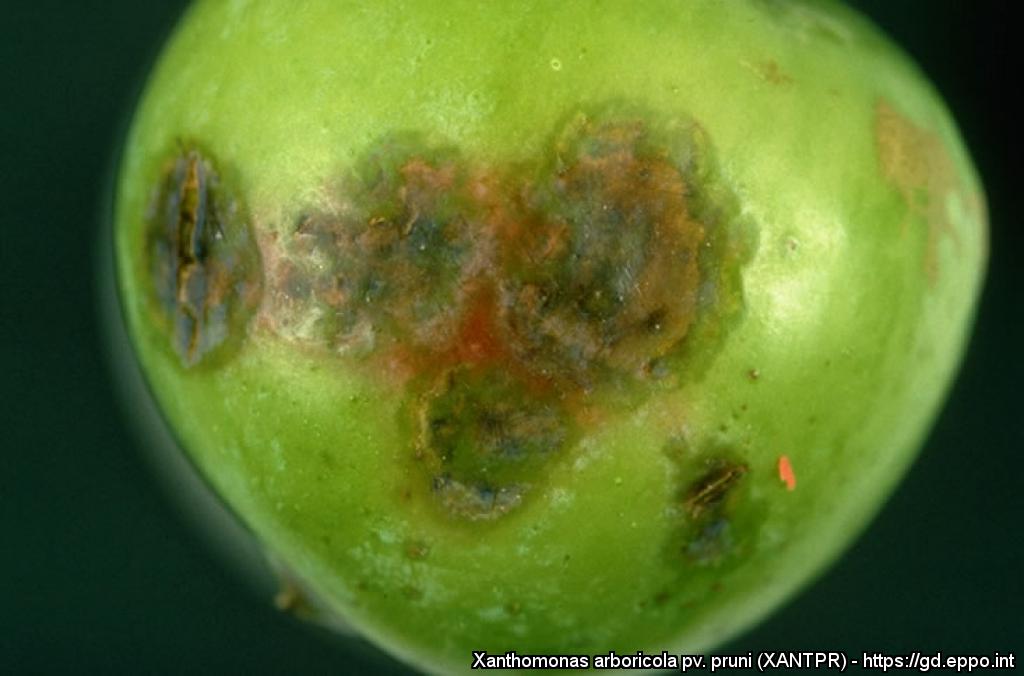
Detail of spots on a plum fruit (P. domestica cv. Black Diamond).
Courtesy: U. Mazzucchi, Universita degli Studi, Bologna (IT).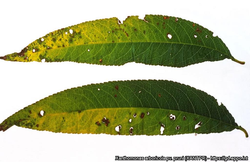
Peach (P. persica) leaves with lesions and shot holes.
Courtesy: U. Mazzucchi, Universita degli Studi, Bologna (IT).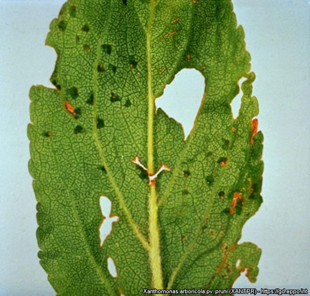
Angular, watersoaked spots on a plum leaf (P. domestica cv. Calita).
Courtesy: U. Mazzucchi, Universita degli Studi, Bologna (IT).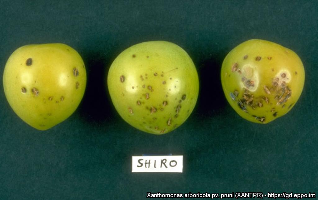
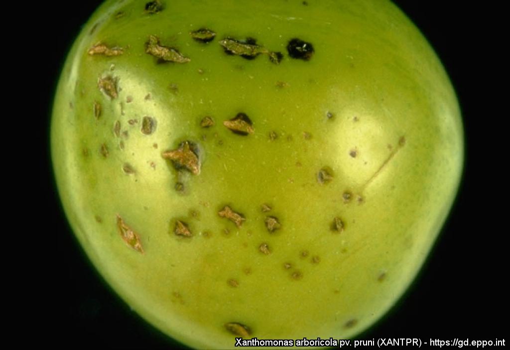
Plum fruits (P. domestica cv. Shiro) with numerous spots.
Courtesy: U. Mazzucchi, Universita degli Studi, Bologna (IT).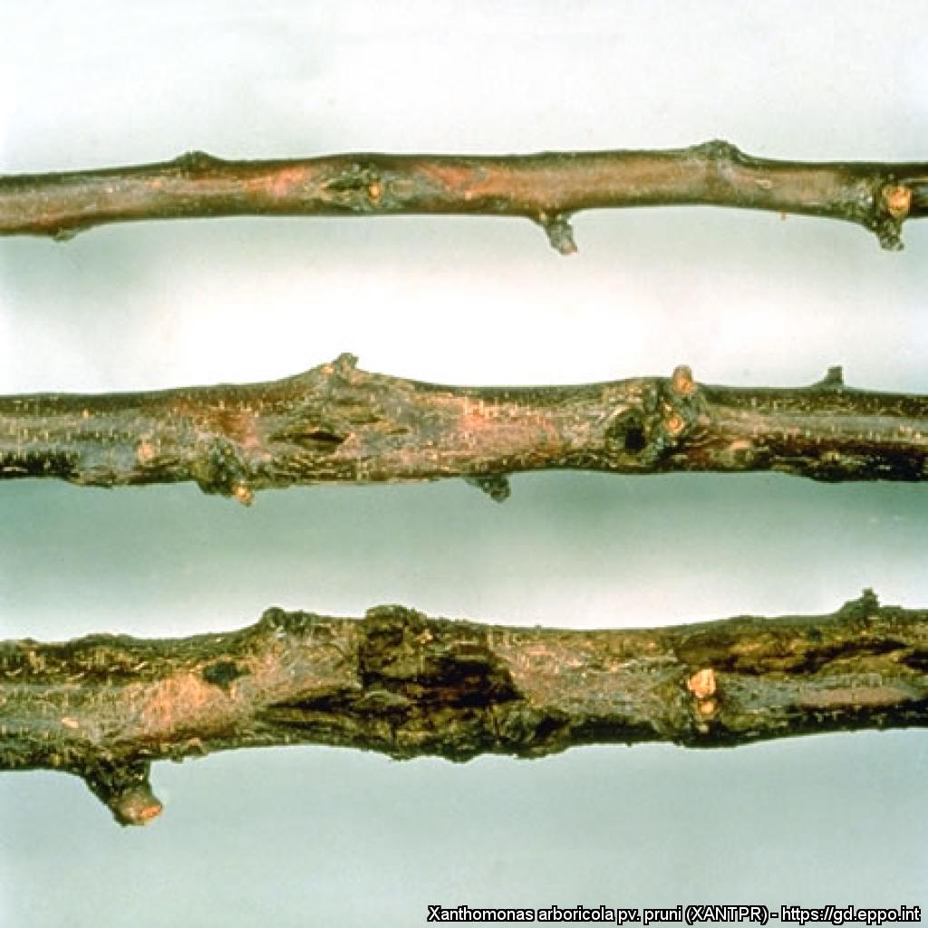
Cankers on three plum branches (P. domestica cv. Calita).
Courtesy: U. Mazzucchi, Universita degli Studi, Bologna (IT).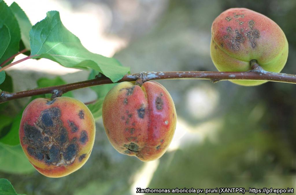
Symptoms on apricots
Courtesy: Miguel Cambra Álvarez (CPV-Government of Aragón, Spain)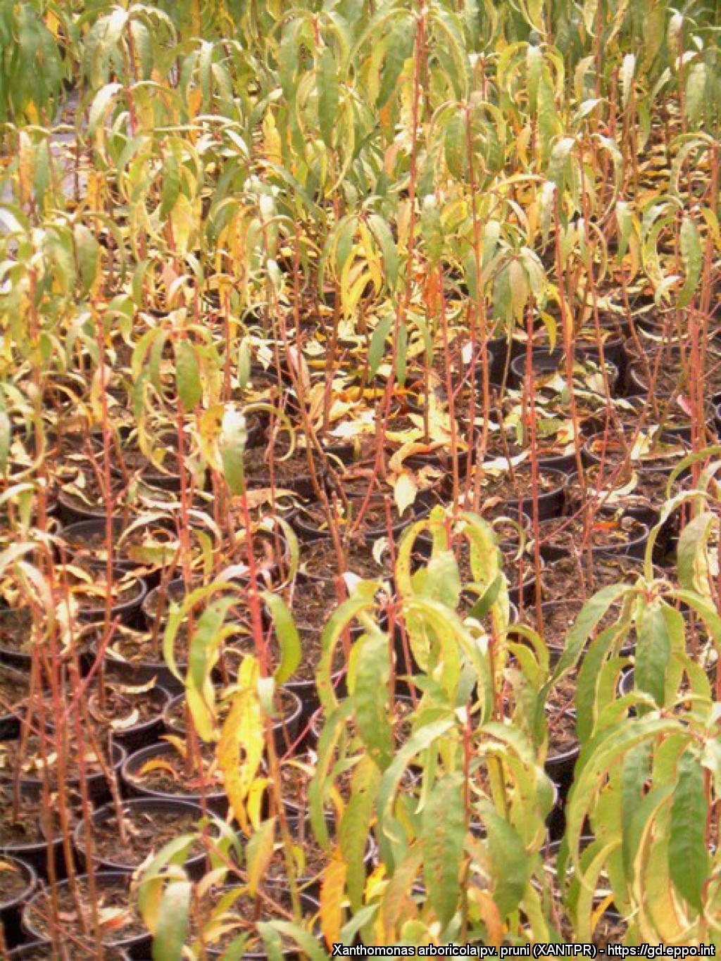
Peach plantlets showing chlorosis of the leaves and heavy defoliation
Courtesy: Plant Health Service, Valencian Government (Spain)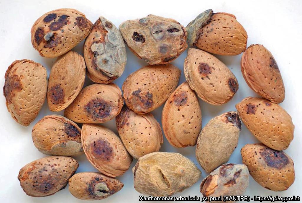
On almonds, in some cases, circular dark spots are observed on the endocarp, which can even affect the nut
Courtesy: Miguel Cambra Álvarez (CPV-Government of Aragón, Spain)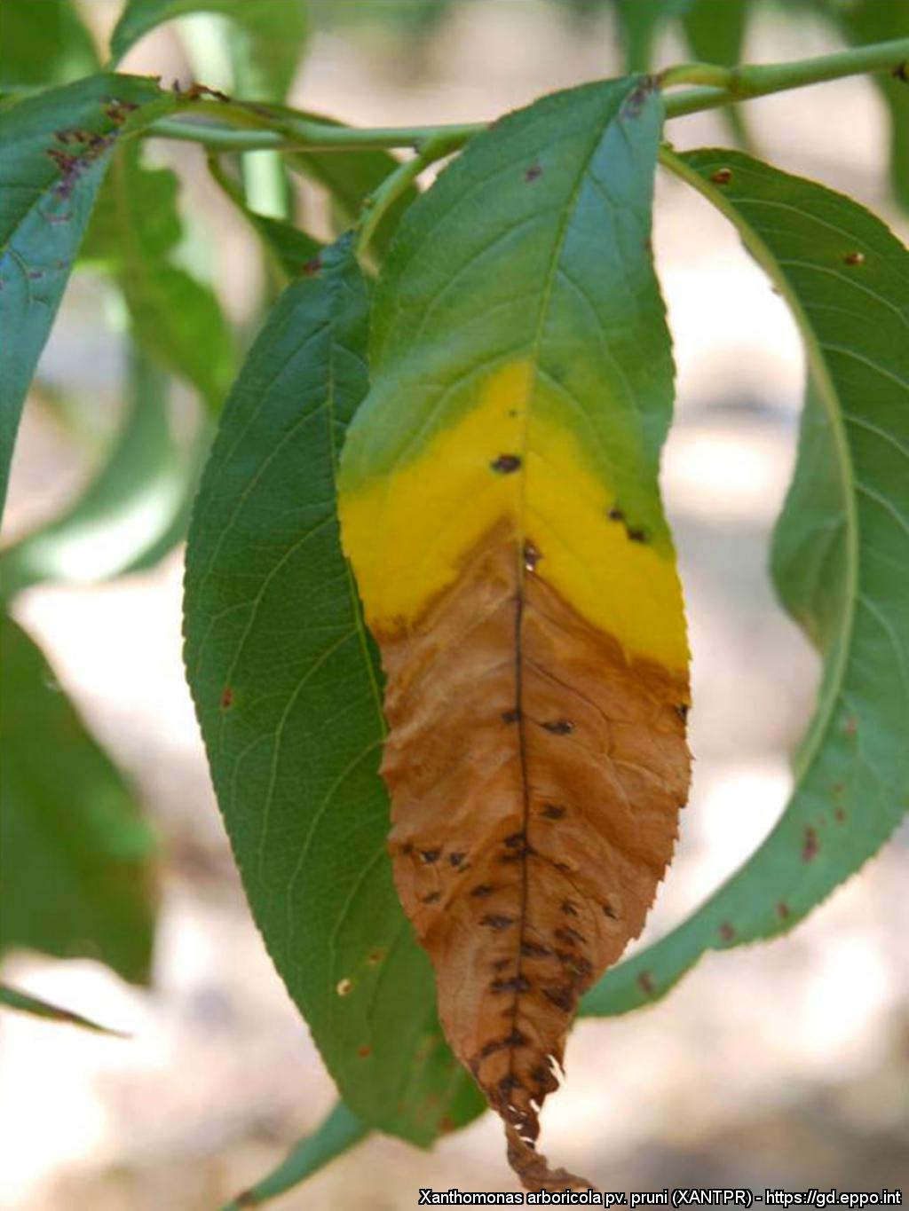
Typical three band colour (green-yellow-brown) symptoms on peach leaves
Courtesy: Miguel Cambra Álvarez (CPV-Government of Aragón, Spain)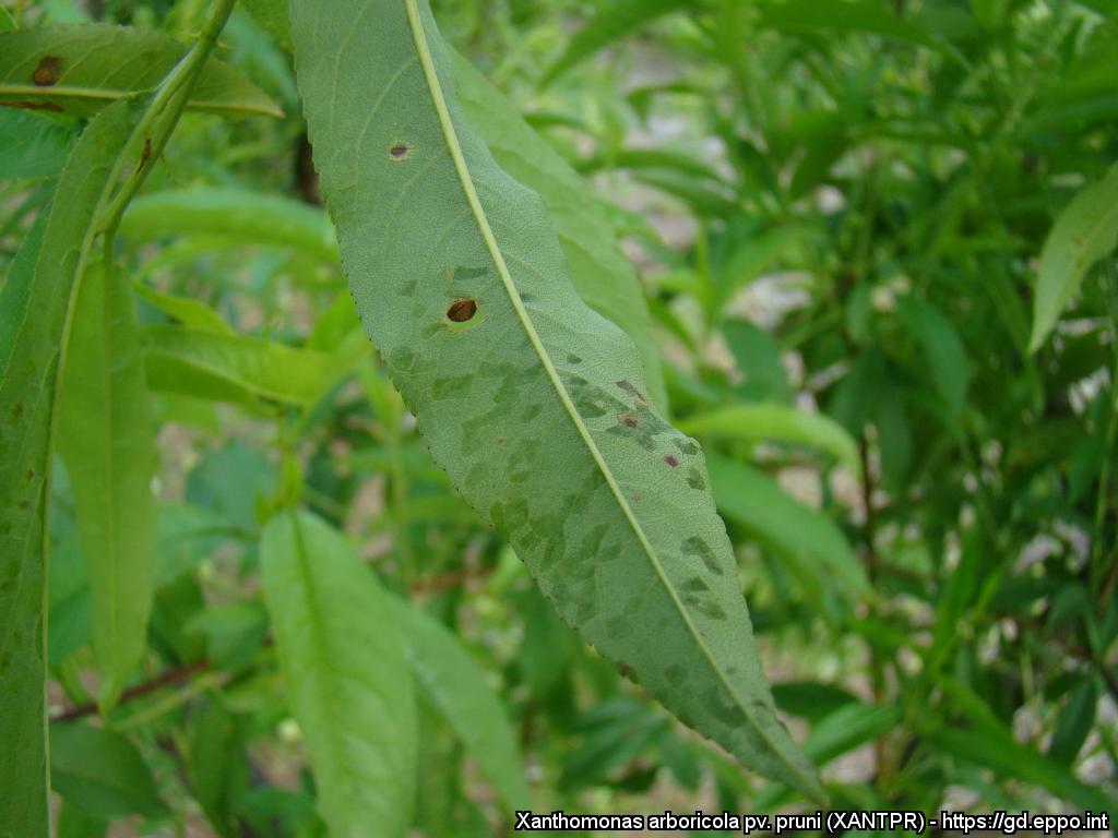
First stage of leaf infection as small, water-soaked lesions on a peach leaf
Courtesy: Miguel Cambra Álvarez (CPV-Government of Aragón, Spain)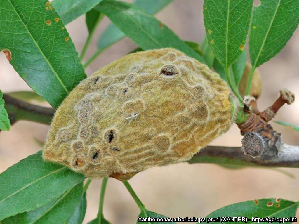
On almonds, when the mesocarp dehydrates, the sunken lesions become raised
Courtesy: Miguel Cambra Álvarez (CPV-Government of Aragón, Spain)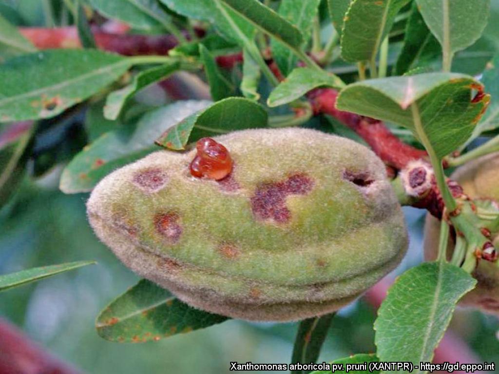
On almonds, infected fruits initially display sunken, corky lesions, oozing gum that streams or clumps
Courtesy: Miguel Cambra Álvarez (CPV-Government of Aragón, Spain)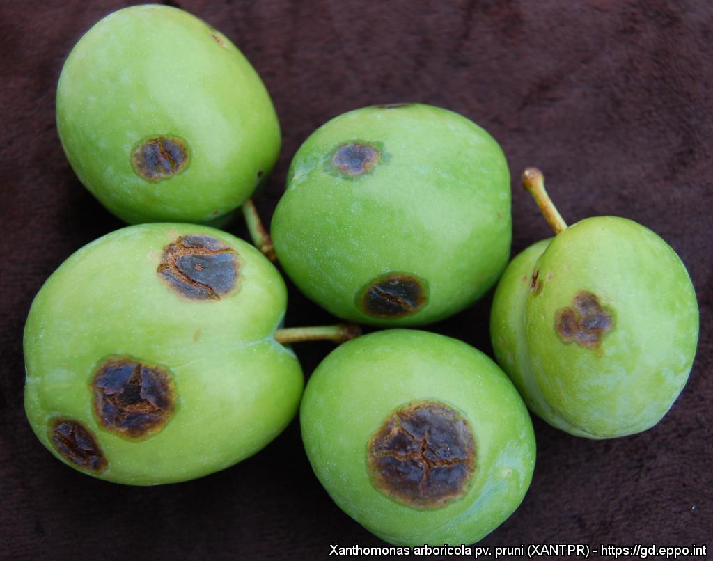
Large necrotic cracking spots on immature fruits of Japanese plum cv. Golden plum. The water-soaked tissue surrounding the necrotic area can easily be observed
Courtesy: Remedios Santiago Merino (LSV-Junta de Extremadura, Spain)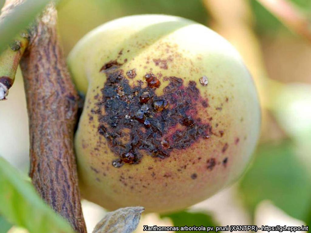
Gum flow from bacterial wounds on a peach
Courtesy: Miguel Cambra Álvarez (CPV-Government of Aragón, Spain)
Almond mummies
Courtesy: Miguel Cambra Álvarez (CPV-Government of Aragón, Spain)
Infected almonds prematurely drop
Courtesy: Miguel Cambra Álvarez (CPV-Government of Aragón, Spain)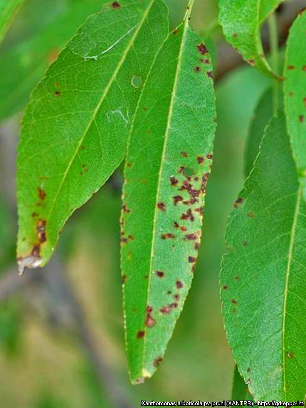
Angular and dark lesions with the chlorotic surrounding tissue on almond leaves
Courtesy: Miguel Cambra Álvarez (CPV-Government of Aragón, Spain)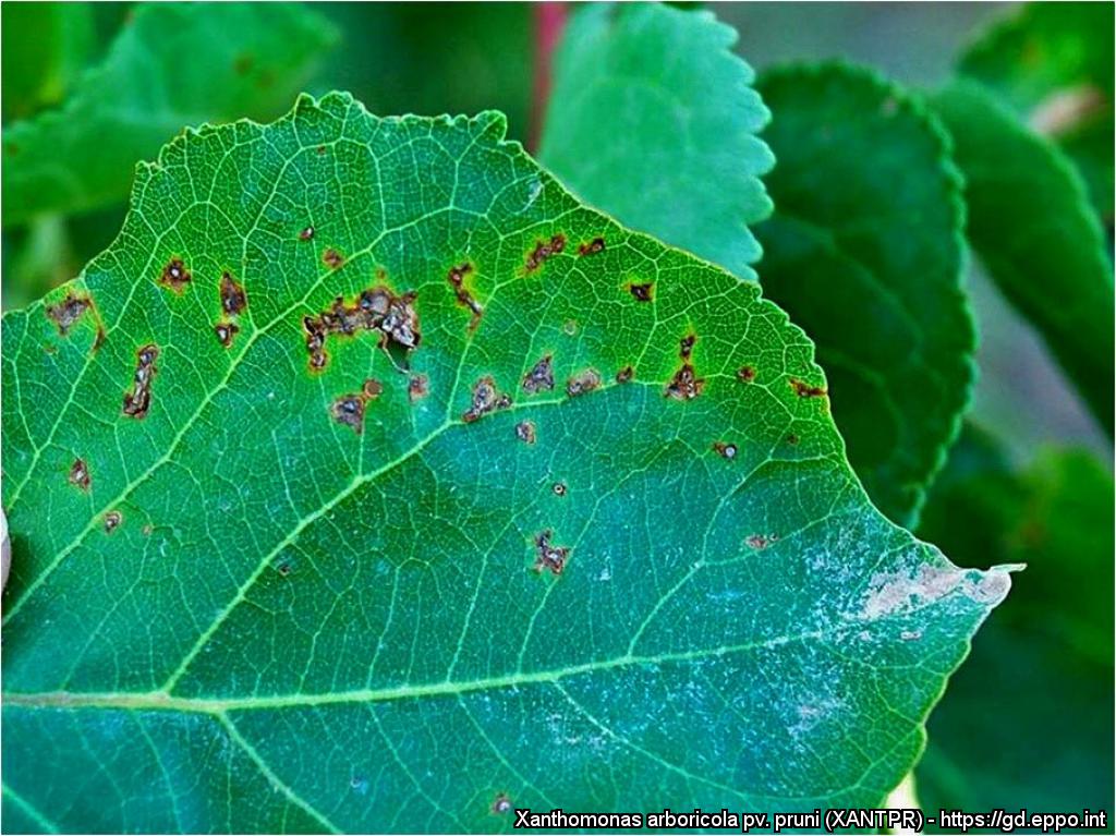
Angular and dark lesions with the chlorotic surrounding tissue on apricot leaf
Courtesy: Miguel Cambra Álvarez (CPV-Government of Aragón, Spain)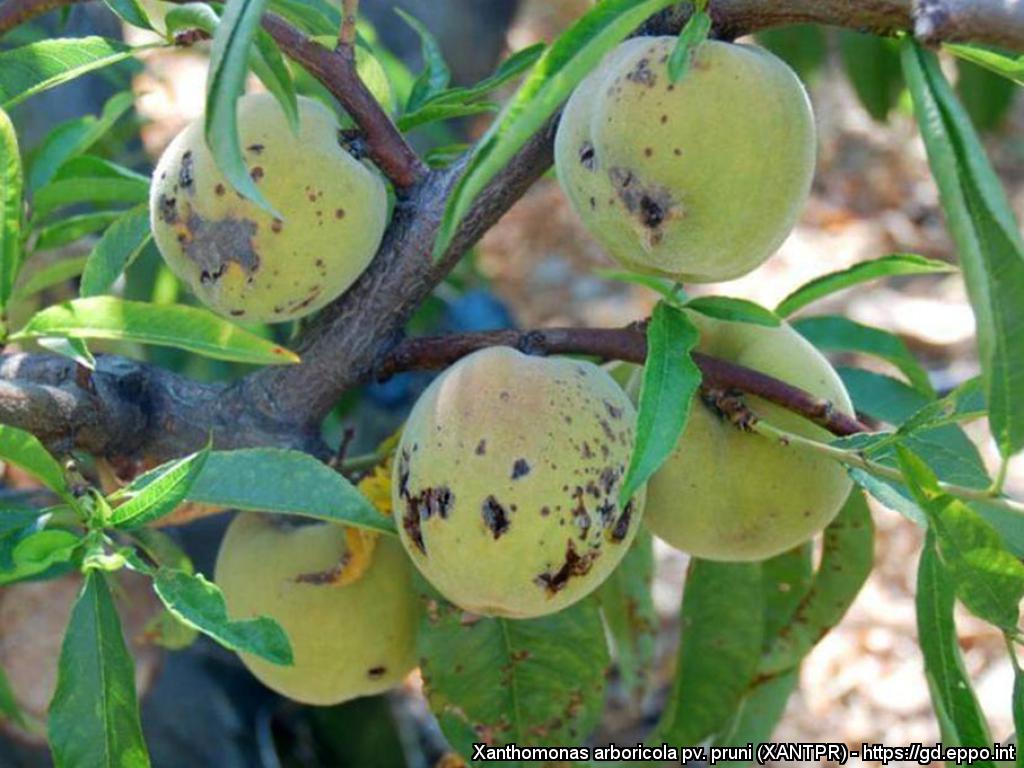
Pitting and cracking occur in the vicinity of the spots, on peaches
Courtesy: Miguel Cambra Álvarez (CPV-Government of Aragón, Spain)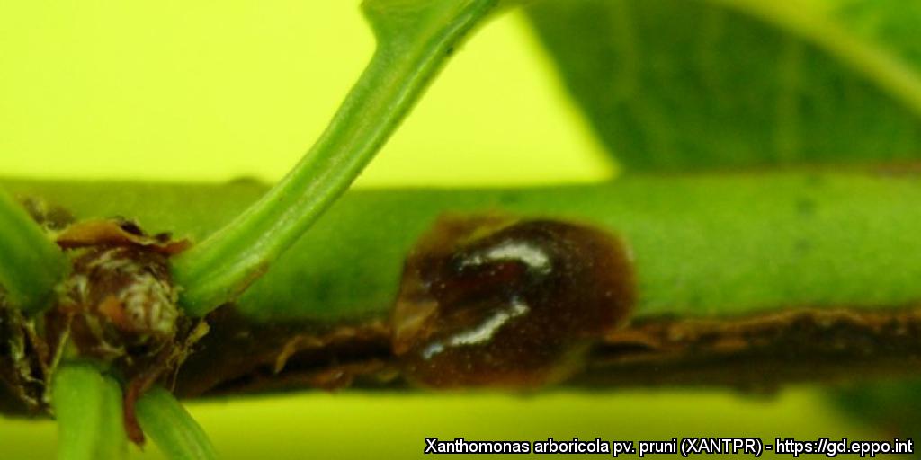
Twig cankers with gum on Prunus dulcis cv. Marta (close-up)
Courtesy: Montserrat Roselló Pérez (LDF-Valencian Government, Spain)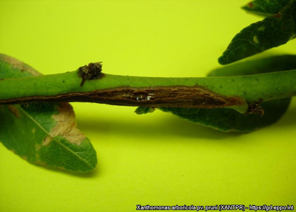
Twig cankers with gum on Prunus dulcis cv. Marta
Courtesy: Montserrat Roselló Pérez (LDF-Valencian Government, Spain)
Tip and twig lesions on young peach shoots
Courtesy: Miguel Cambra Álvarez (CPV-Government of Aragón, Spain)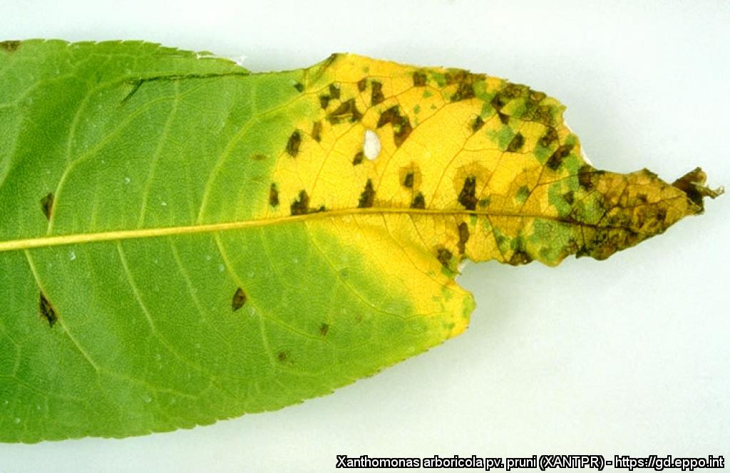
Detail of a peach leaf (P. persica cv. Maria Serena).
Courtesy: U. Mazzucchi, Universita degli Studi, Bologna (IT).