Xylotrechus chinensis(XYLOCH)
Photos
For publication in journals, books or magazines, permission should be obtained from the original photographers with a copy to EPPO.
Adult Xylotrechus chinensis detected in the region of Heraklion (Kriti, Greece)
Courtesy: Leivadara et al. 2018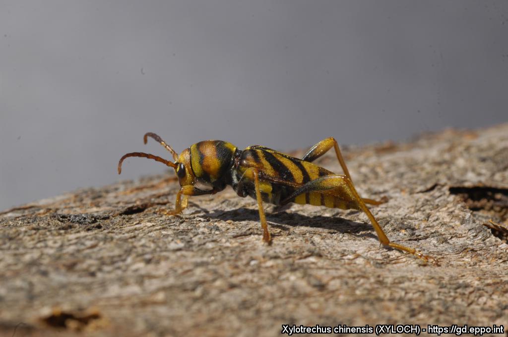
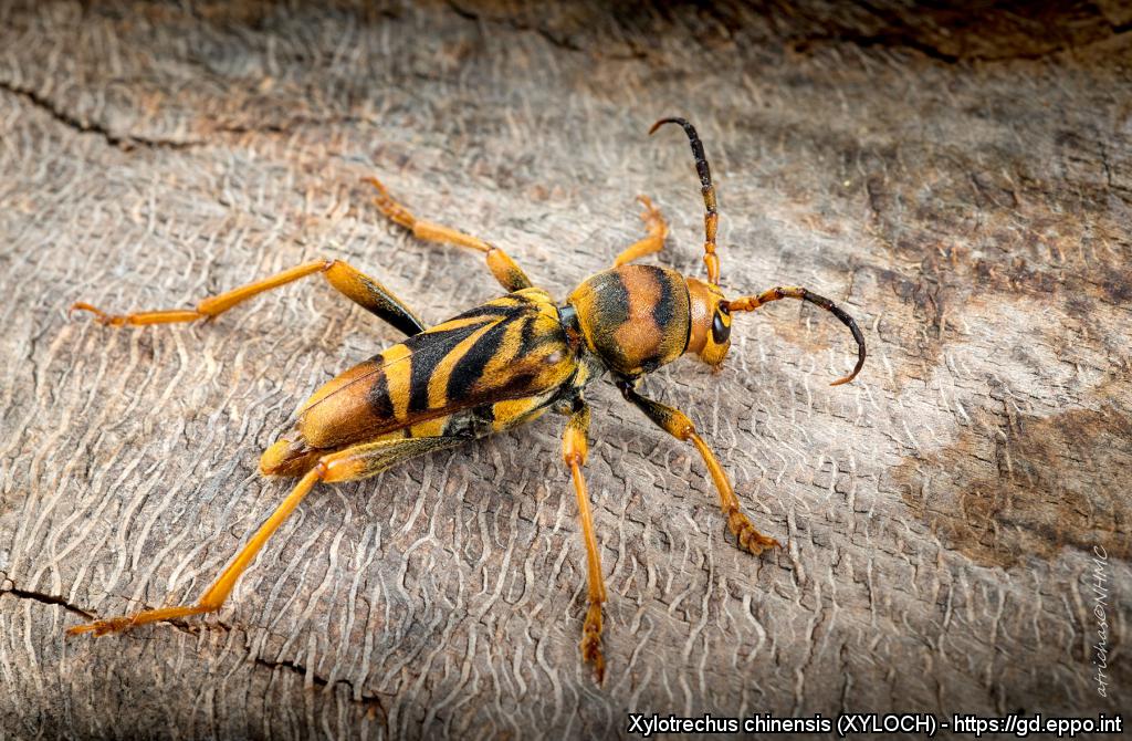
Adult female X. chinensis
Courtesy: Victor Sarto i Monteys Institute of Environmental Science and Technology (ICTA), Spain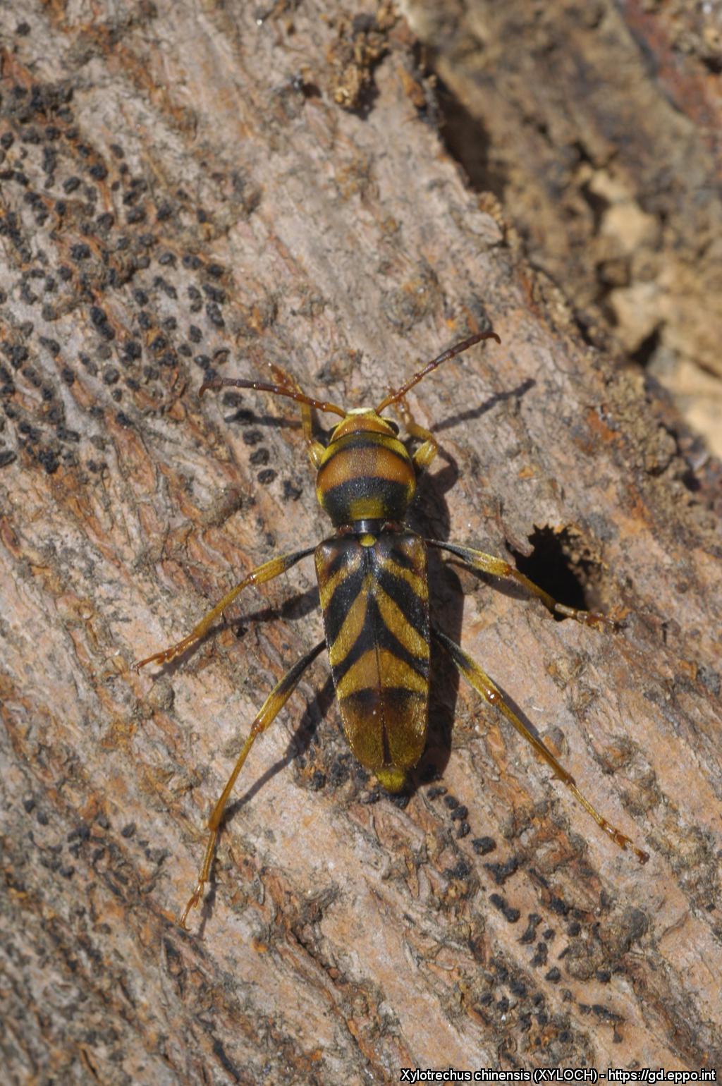
Adult female X. chinensis
Courtesy: Victor Sarto i Monteys Institute of Environmental Science and Technology (ICTA), Spain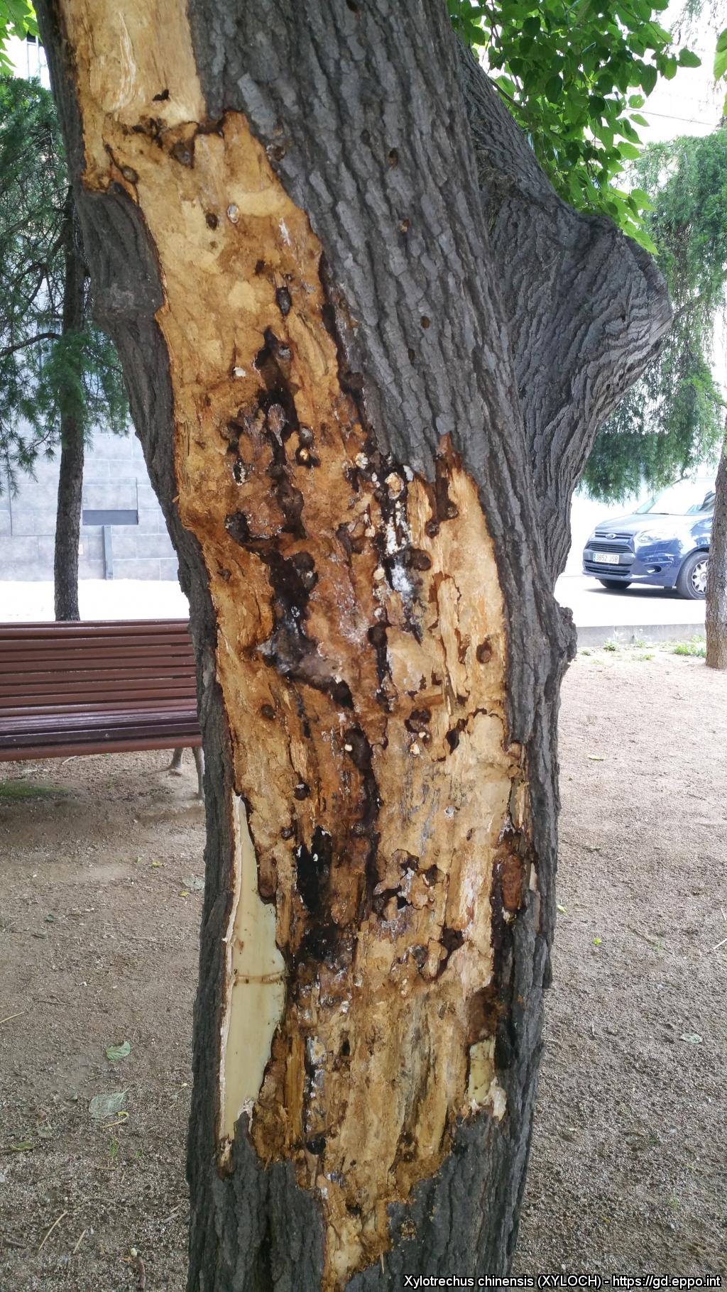
Larval galleries of X. chinensis on Morus trees (bark removed)
Courtesy: Àngels Blanquez (Ripollet municipality, Spain)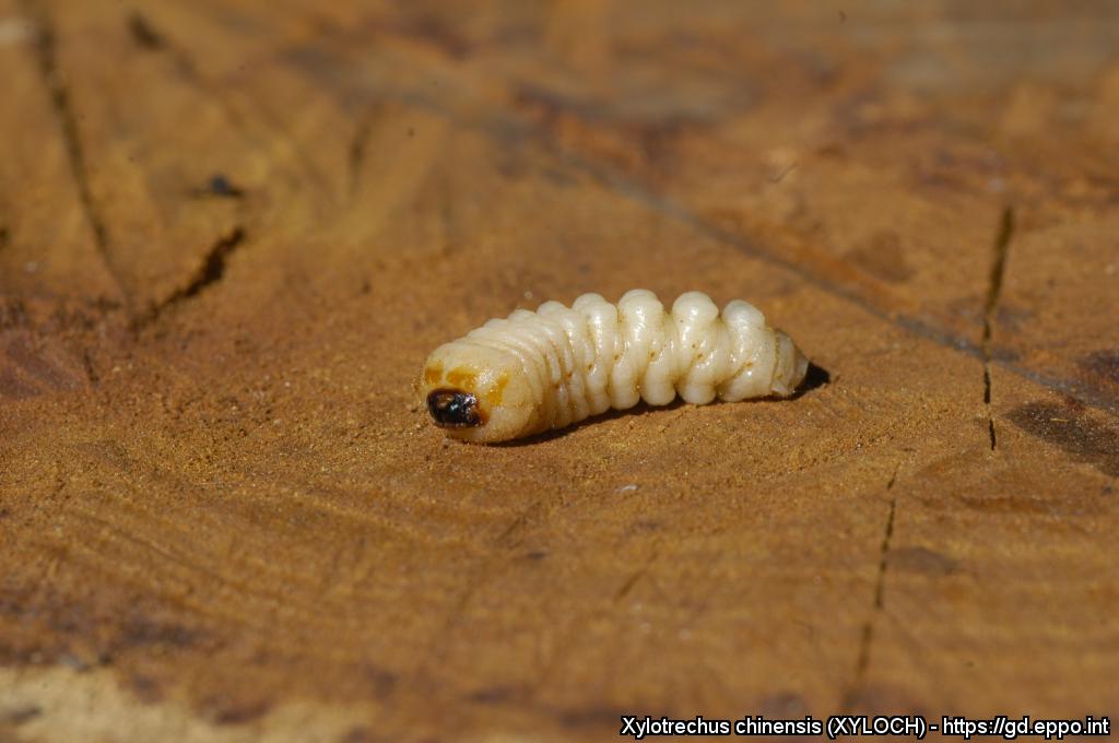
Last instar larva of X. chinensis (extracted from its cavity)
Courtesy: Victor Sarto i Monteys Institute of Environmental Science and Technology (ICTA), Spain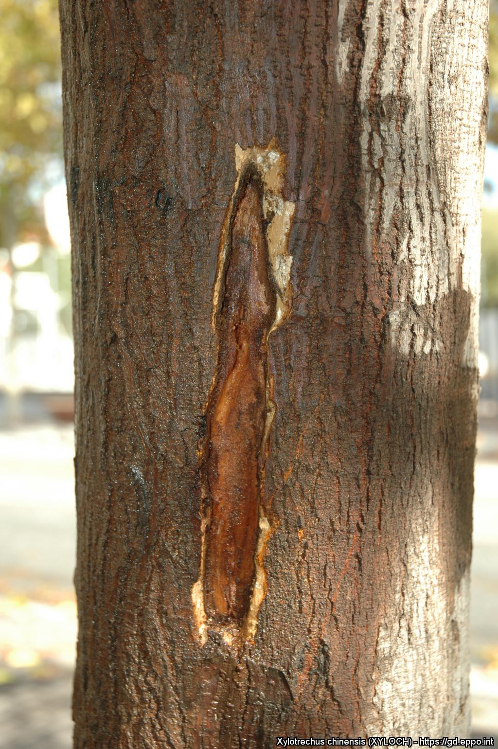
X. chinensis larva within its feeding cavity in the phloem of a mulberyy tree trunk (bark removed)
Courtesy: Victor Sarto i Monteys Institute of Environmental Science and Technology (ICTA), Spain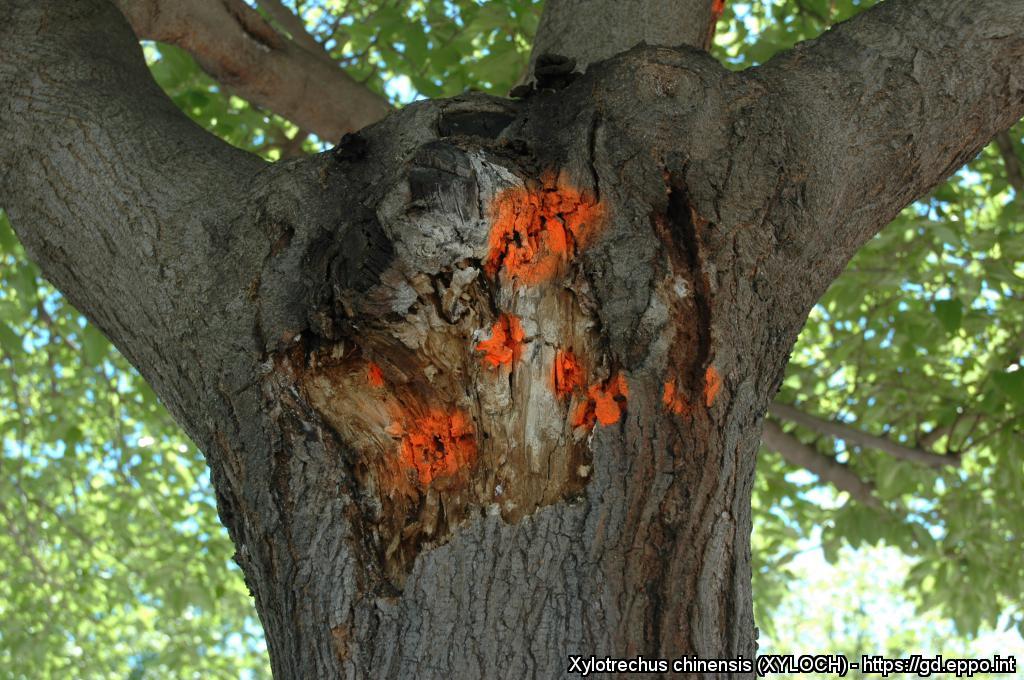
Exposed xylem sapwood beginning to dry and crack after heavy damage by X. chinensis larvae
Courtesy: Victor Sarto i Monteys Institute of Environmental Science and Technology (ICTA), Spain
Head of male adult X. chinensis (note the widely separated antennae)
Courtesy: Victor Sarto i Monteys Institute of Environmental Science and Technology (ICTA), Spain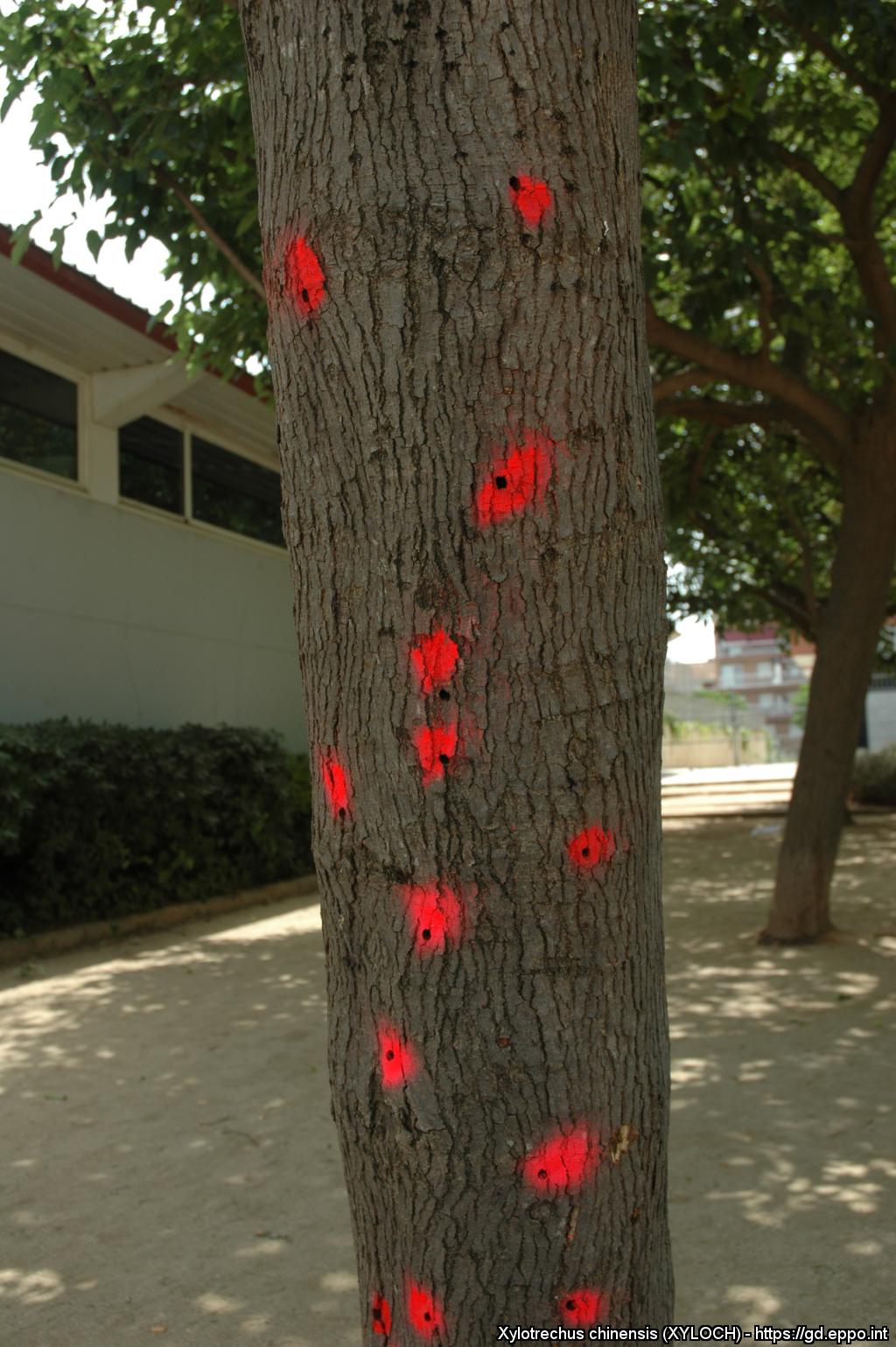
Adult emergence holes of X. chinensis in an heavily infested mulberry tree
Courtesy: Victor Sarto i Monteys Institute of Environmental Science and Technology (ICTA), Spain
Adult emergence holes of X. chinensis in a heavily infested mulberry tree
Courtesy: Glòria Torras i Tutusaus (Ajuntament de Barberà del Vallès, Spain)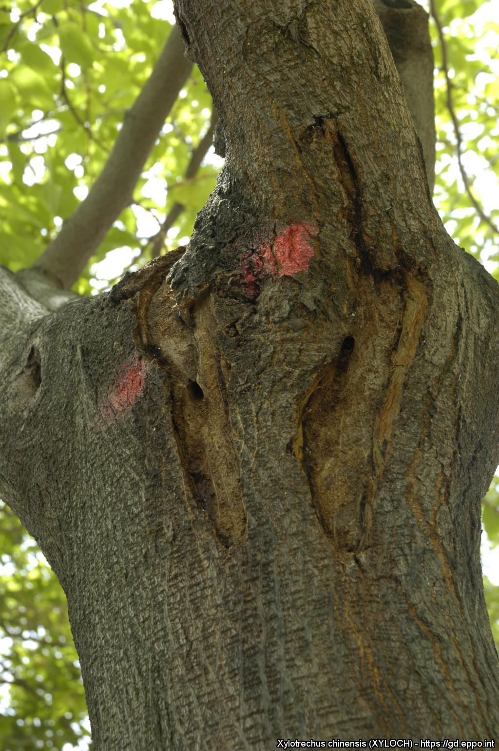
Mulberry trunk with detached periderm showing consumed and dead phloem as well as adult emergence holes
Courtesy: Victor Sarto i Monteys Institute of Environmental Science and Technology (ICTA), Spain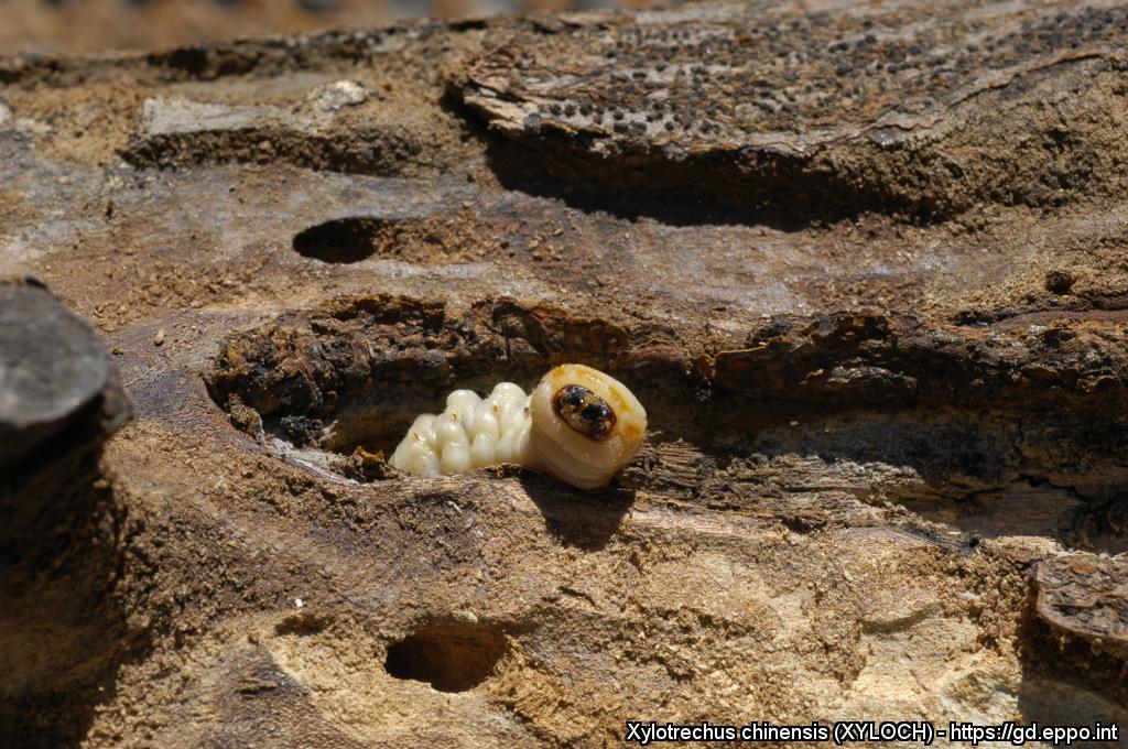
Larva of X. chinensis in a mulberry trunk
Courtesy: Victor Sarto i Monteys Institute of Environmental Science and Technology (ICTA), Spain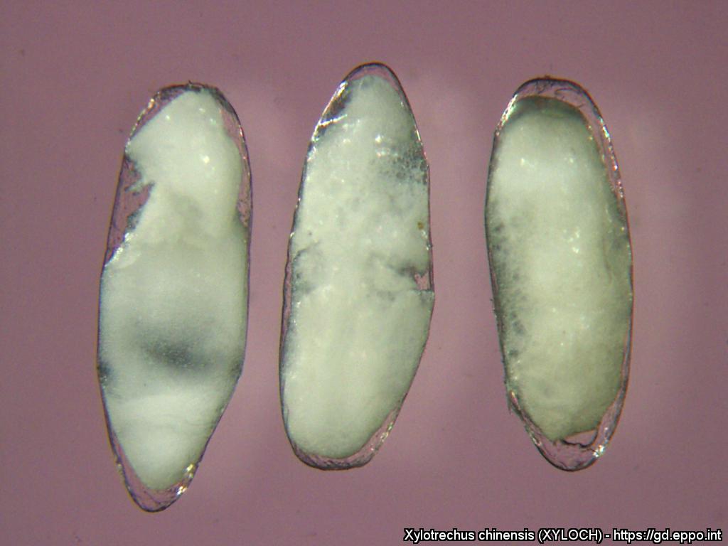
Eggs (unfertilized) of X. chinensis showing the oocyte mass protected by a transparent chorion
Courtesy: Victor Sarto i Monteys Institute of Environmental Science and Technology (ICTA), Spain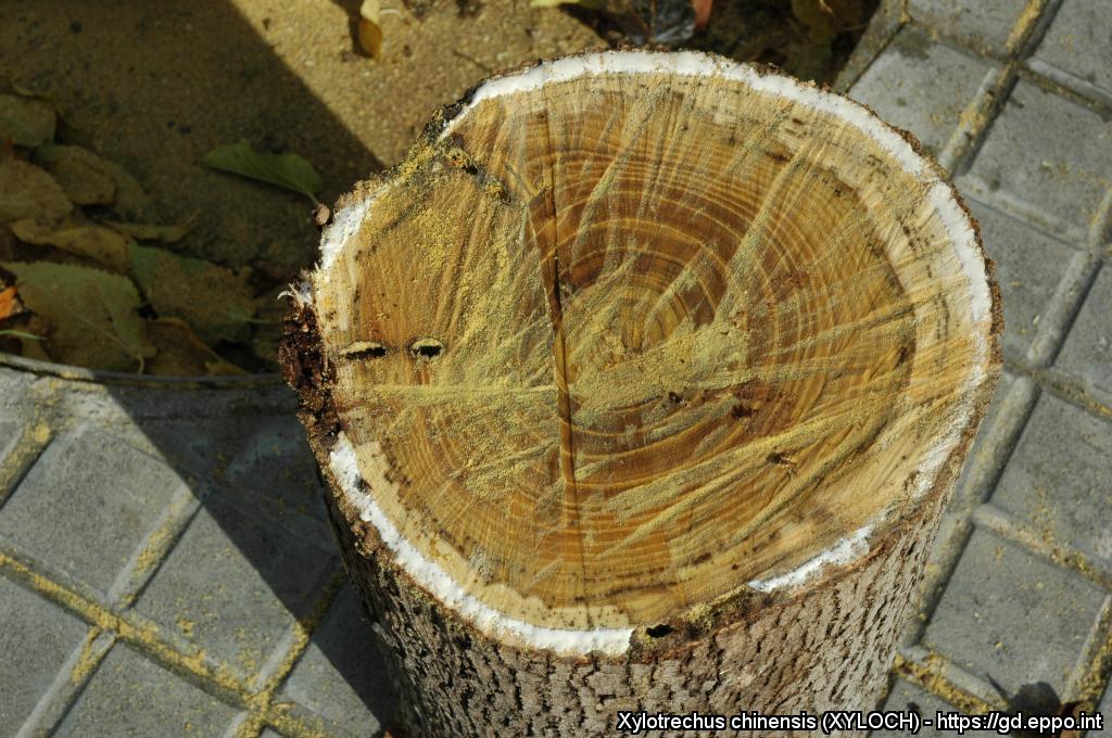
X. chinensis phloem and xylem larval cavities. Note white latex exuding in mulberry phloem after cutting after cutting the trunk, excepting the sections consumed by the beetle's larvae
Courtesy: Victor Sarto i Monteys Institute of Environmental Science and Technology (ICTA), Spain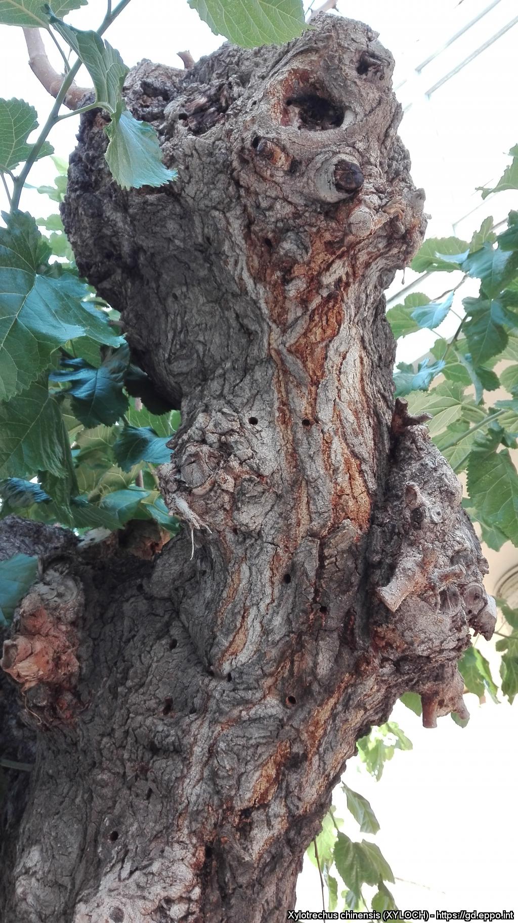
Exit holes of adult Xylotrechus chinensis on mulberry trunks near the harbour fo Heraklion (Kriti, Greece)
Courtesy: Leivadara et al. 2018